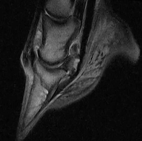
In 2018, Equine Practice introduced the service of Magnetic Resonance Imaging (MRI) for the
diagnosis of the most common orthopaedic conditions of the equine athlete. The examination
is carried out with the use of a system specifically designed by ESAOTE, a company leader in
MRI, which has been involved in the veterinary field for a long time, in the imaging of the
distal limb of the horse. The O-Scan Equine, acquired by the hospital, has already been
adopted by many distinguished private and college institutions in Europe and North America
for its performance comparable to the image quality of high field systems. The system covers
imaging of the foot, pastern, fetlock, high suspensory, carpus and tarsus regions of the equine
limb.
The examination with the O-Scan Equine is performed in general anaesthesia, like the high
field systems that guarantee a better quality of the images in relation to their performance
and the absence of movement during the acquisition that can compromise the quality of the
examination and its duration, in particular, for the fetlock and regions above it as can happen
in the standing system. The choice of a system that requires a general anaesthesia to perform
an MRI has never represented a great concern for us, considering the great experience
acquired by our anaesthesiologist staff in over 30 years of practice and many thousands of
procedures performed.
The examination can be reserved and performed in a very short time with the help of the
treating veterinarian who must inform the colleagues of the clinic on the previous clinical and
instrumental examinations performed on the horse to address, in the best way possible, the
area to be examined. The previous history improves the interpretation of the findings that is
guaranteed by the report of Dr. Silvia Rabba, a specialist in veterinary imaging and eventually,
when required, by the clinical interpretation and management suggested by Dr. Fernando Canonici, a well distinguished veterinary surgeon of great experience in the orthopaedic
conditions of the equine athlete.


